Our Medical Technology
Eaglet Eye – Eye Surface Profiler for Scleral Profilometry
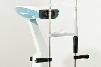
The Eaglet-Eye ESP offers more features than any previous profiler. Our revolutionary topographer is the first to offer profiling, not only of the cornea, but also right across the limbus and over a large portion of the sclera.
Click here to learn more about Scleral Profilometry
Corneal Mapping
Corneal topography, also known as photokeratoscopy or videokeratography, is a non-invasive medical imaging technique for mapping the surface curvature of the cornea, the outer structure of the eye. Since the cornea is normally responsible for some 70% of the eye’s refractive power, its topography is of critical importance in determining the quality of vision.
The three-dimensional map is therefore a valuable aid to the examining ophthalmologist or optometrist and can assist in the diagnosis and treatment of a number of conditions; in planning refractive surgery such as LASIK and evaluation of its results; or in assessing the fit of contact lenses. A development of keratoscopy, corneal topography extends the measurement range from the four points a few millimeters apart that is offered by keratometry to a grid of thousands of points covering the entire cornea. The procedure is carried out in seconds and is completely painless.
Special thanks to the EyeGlass Guide, for informational material that aided in the creation of this website. Visit the EyeGlass Guide today!
Digital Retinal Imaging & OCT Scans
We use cutting-edge digital imaging technology to assess your eyes. Many eye diseases, if detected at an early stage, can be treated successfully without total loss of vision. Your retinal Images will be stored electronically. This gives the eye doctor a permanent record of the condition and state of your retina.
This is very important in assisting your Optometrist to detect and measure any changes to your retina each time you get your eyes examined, as many eye conditions, such as glaucoma, diabetic retinopathy and macular degeneration are diagnosed by detecting changes over time.
The advantages of digital imaging include:
- Quick, safe, non-invasive and painless
- Provides detailed images of your retina and sub-surface of your eyes
- Provides instant, direct imaging of the form and structure of eye tissue
- Image resolution is extremely high quality
- Uses eye-safe near-infra-red light
- No patient prep required
Digital Retinal Imaging
Digital Retinal Imaging allows your eye doctor to evaluate the health of the back of your eye, the retina. It is critical to confirm the health of the retina, optic nerve and other retinal structures. The digital camera snaps a high-resolution digital picture of your retina. This picture clearly shows the health of your eyes and is used as a baseline to track any changes in your eyes in future eye examinations.
Optical Coherence Tomography (OCT)
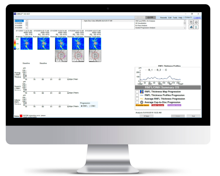
An Optical Coherence Tomography scan (commonly referred to as an OCT scan) is the latest advancement in imaging technology. Similar to ultrasound, this diagnostic technique employs light rather than sound waves to achieve higher resolution pictures of the structural layers of the back of the eye.
At 100,000 scans per second, Zeiss Cirrus 6000 enables Dr. Raghu to image a larger field of view up to 12mm in a single scan. It also captures high-definition (HD) OCT and OCT Angiography (OCTA) scans, revealing the finer microvascular details of the retina. These images allow us to track macular changes, glaucoma, epithelial thickness and retinal health.
An OCT scan is a noninvasive, painless test. It is performed in about 10 minutes right in our office. Feel free to contact our office to inquire about an OCT at your next appointment.
Visual Field Testing
A visual field test measures how much ‘side’ vision you have. It is a straightforward test, painless, and does not involve eye drops. Essentially lights are flashed on, and you have to press a button whenever you see the light. Your head is kept still and you have to place your chin on a chin rest. The lights are bright or dim at different stages of the test. Some of the flashes are purely to check you are concentrating.
Each eye is tested separately and the entire test takes 15-45 minutes. Your optometrist may ask only for a driving licence visual field test, which takes 5-10 minutes. If you have just asked for a driving test or the clinic doctor advised you have one, you will be informed of the result by the clinic doctor, in writing, in a few weeks.
Normally the test is carried out by a computerised machine, called a Humphrey. Occasionally the manual test has to be used, a Goldman. For each test you have to look at a central point then press a buzzer each time you see the light.
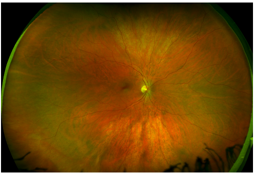
OPTOS Retinal Exam
Annual eye exams are vital to maintaining your vision and overall health.
Our Optos machine produces high resolution images of approximately 82% or 200◦ of the retina. These images provide a bigger picture and more clinical information which facilitates the early detection, management and effective treatment of disorders and diseases evidenced in the retina such as retinal detachments and tears, glaucoma, diabetic retinopathy and age-related macular degeneration
Many eye problems can develop without you knowing. You may not even notice any change in your sight.
An optomap® Retinal Exam provides:
- A scan to show a healthy eye or detect disease.
- A view of the retina, giving your doctor a more detailed view than he/she can get by other means.
- The opportunity for you to view and discuss the optomap® image of your eye with your doctor at the time of your exam.
- A permanent record for your file, which allows us to view your images each year to look for changes.
The optomap® Retinal Exam is fast, easy, and comfortable for all ages. To have the exam, you simply look into the device one eye at a time and you will see a comfortable flash of light to let you know the image of your retina has been taken. The optomap® image is shown immediately on a computer screen so we can review it with you.
Please schedule your optomap® Retinal Exam today!
For more information on the optomap® Retinal Exam, go to the Optos website.
LipiFlow & Meibography
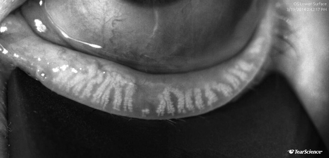 In about 60 seconds the LipiScan Dynamic Meibomian Imager allows us to take images of the glands and lipid-layer thickness to test for dry eye. With these images we can treat the root cause of your dry eye.

Lens Technology
UV Protect Technology moves beyond today’s standards to a higher level of UV protection, which allows clear ZEISS lenses to block harmful UV rays up to 400 nm. This is the same standard of UV protection premium sunglasses provide.
DuraVision BlueProtect provides more visual comfort and greater safety. Modern light sources and displays emit a disproportionately high amount of blue light. While the human body requires a certain amount of blue light to control the sleep/wake cycle, too much of the ‘wrong’ kind of blue light can pose a health risk and cause eye diseases. Lenses with this coating feature a special filter which ensures comfortable vision even when there is a high level of blue light present.
Progressive Lens designs enable us to produce a pair of progressive glasses optimized for your particular vision needs and featuring unprecedented quality. Cutting-edge computer technology assists in measuring visual performance and fitting the lenses to your frames. The near, intermediate and distance zones are all incorporated into one lens with smooth transitions. These lenses are tailor-made for your facial shape, your lifestyle, your career and your visual habits. Individual and one-of-a kind – just like you.
The Digital Lens design offers the perfect remedy to digital eye strain by enabling fast, comfortable focusing thanks a specially optimized near zone – no matter where we look. These lenses are ideal for individuals who not only use digital devices, but also spend time reading books and magazines.
The Scleral Lens design allows people who would normally be unable to wear contacts the ability to wear them.
The Overnight Vision Correction Lens is put in at night and corrects your vision while you sleep.